
Oral and Maxillofacial Radiology
Oral and Maxillofacial Radiology is a specialty recognized by the Canadian Dental Association. Oral and Maxillofacial Radiology is the branch and specialty of dentistry concerned with the prescription, production, and interpretation of diagnostic images for the diagnosis and management of diseases and disorders of the craniofacial complex.
Examination Information
The Oral and Maxillofacial Radiology examination will be held in person at a single location in Toronto, Ontario. The exact location of the examination site will be communicated after the closure of the 2026 Fellowship Examination application window.
| Element | Structure |
|---|---|
| Part 1: Clinical | 4 cases (80 minutes) |
| Part 2: OSCE | 8 stations x 20 minutes (2 hours 40 minutes) |
Part 1 (Clinical)
This section consists of four structured written cases (80 minutes total). Media may include DICOM datasets viewable in Anatomage Invivo, PowerPoint slide decks, or videos playable with VLC. The patient’s age and sex will be provided. Candidates are expected to produce a concise radiology report for each case.
| Format | Each session consists structured written cases. |
| Candidate Role | Candidates are expected to write a Radiology report for each case as they would in practice. |
Part 2 (OSCE)
An Objective Structured Clinical Examination (OSCE) is a practical assessment designed to evaluate the clinical competence of candidates in a structured and systematic way. The format of the examination allows candidates to be assessed by multiple examiners across different stations, ensuring a comprehensive evaluation of their skills. As a result, candidates may encounter an Examiner from their own dental specialty training program. Significant conflicts, such as being examined by a program director, are identified and avoided when scheduling the examination. Each station presents clinical scenarios or radiographic cases that test candidates’ ability to interpret imaging, diagnose, and apply radiologic principles in real-world situations, providing a consistent and fair evaluation of their diagnostic reasoning, image analysis, and clinical decision-making skills.
| Format | Each session consists of a series of structured clinical cases designed to reflect real-world oral and maxillofacial radiology practice. |
| Assessment | Performance is evaluated using the Global Rating Scale (GRS), which emphasizes judgment, reasoning, and communication across multiple domains. |
| Examiner Role | Examiners are responsible for pacing the examination and guiding the discussion. |
| Candidate Role | Candidates are expected to approach each case as they would in practice: by gathering and interpreting information, formulating a diagnosis, developing and justifying management options, and communicating their decisions clearly. Candidates are encouraged to think aloud, explain their reasoning, and communicate their approach as they would in a clinical setting. |
Examination Blueprint
| Domain | Weight (%) |
|---|---|
| Diagnostic Images and Imaging Modalities | 5 – 15% |
| Pertinent Radiological Observations | 30 – 40% |
| Interpretation of Imaging Findings and Diagnosis | 30 – 40% |
| Recommendations for Case Management | 15 – 25% |
Blueprint
Oral and Maxillofacial Radiology

Preparation
Preparation should focus on applied reasoning and communication rather than memorization. Candidates are encouraged to:
- Practice structured case discussions, individually or with peers.
- Rehearse verbalizing reasoning, including justification of decisions and management of uncertainty.
- Review the example case provided to become familiar with the style of questioning and depth of response expected.
- Practice producing succinct radiology reports and structured pertinent observations from multi-modality datasets under timed conditions.
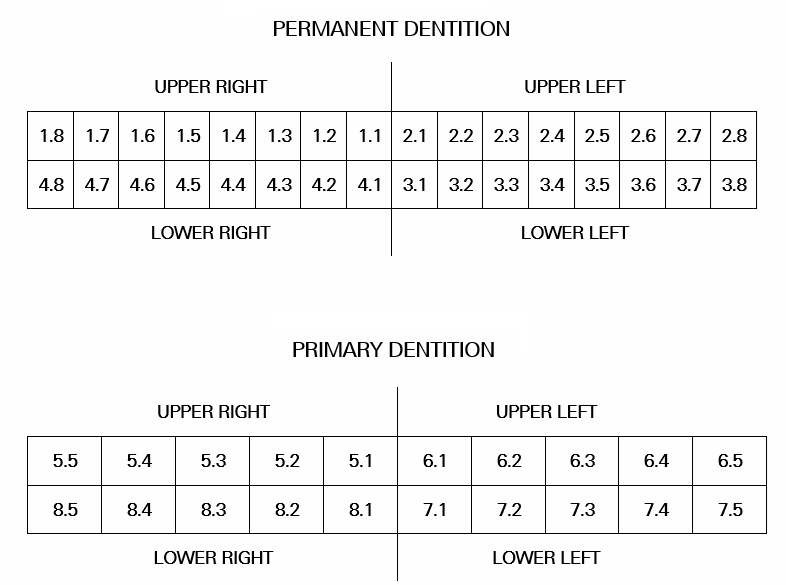
FDI Two-Digit Tooth Numbering Chart
The Fellowship Examination uses the FDI Two-Digit system.
Sample Case
Oral and Maxillofacial Radiology